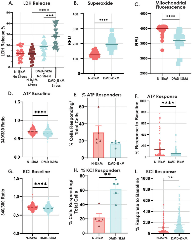
Fig. 2.
DMD-iSkMs have altered stress injury, ROS, mitochondrial and calcium-handling phenotypes compared to control. (A) DMD-iSkMs displayed significantly greater LDH release than N-iSkMs under stressed conditions (P-value<0.0001). (B) Superoxide as an indicator of ROS is increased in DMD-iSkMs (P-value<0.0001). (C) Mitochondrial fluorescence as an indicator of mitochondrial function was decreased in DMD-iSkMs as compared to N-iSkMs (P-value<0.0001). D-F) DMD-iSkMs had a significant decrease in bound calcium (P-value<0.0001), a trending decrease in the percent of ATP responders, and a significant decrease in ATP response compared to baseline as compared to N-iSkMs (P-value<0.0001). (G-I) At baseline, bound calcium was significantly decreased before KCl stimulation in DMD-iSkMs (P-value<0.0001). There was a significant increase in the percent of cells responding (P-value=0.0014) and the response to baseline in DMD-iSkMs after KCl stimulation (P-value<0.0001).

