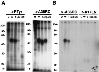
FIG. 3.
SDS-PAGE analysis of tyrosine-phosphorylated proteins from infected cells. BS-C-1 cells were uninfected (U) or were infected with vaccinia virus strain WR (W) or IHDJ (I) or with a recombinant vaccinia virus from which the A33R ORF (ΔA33) or the A36R ORF (ΔA36) had been deleted. Cells were then labeled with H332PO4. (A) Lysates were immunoprecipitated with either the anti-phosphotyrosine antibody PY99 or α-A36RC and were analyzed by SDS-PAGE. (B) An equivalent portion of the material immunoprecipitated by PY99 was eluted from the protein A-coupled antibody with cold phosphotyrosine and a high salt concentration. This postelution fraction was diluted and immunoprecipitated with either α-A36RC or antibody to the N-terminal region of A17L (anti-A17LN). The washed samples were analyzed by SDS-PAGE and autoradiography. The masses (in kilodaltons) and positions of migration of markers are indicated on the left.

