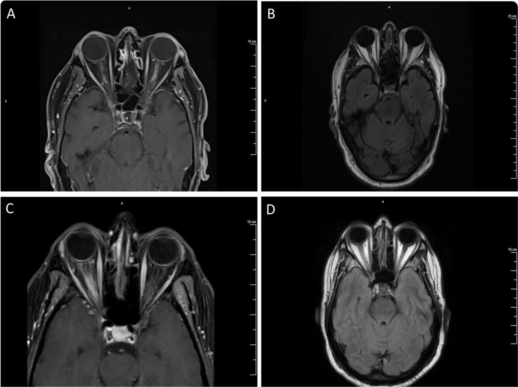
Figure 1. MRI images.

Case 1: Patchy and irregular enhancement involving the intraorbital segments of the bilateral optic nerve, left greater than right (A), with mild fusiform enlargement associated with T2 hyperintensity in the left optic nerve with preservation of surrounding subarachnoid space (B). Case 2: Abnormal enhancement (C) and T2 hyperintensity (D) in the right optic nerve extending from posterior intraorbital segment to canalicular segment with associated surrounding perineural enhancement.
