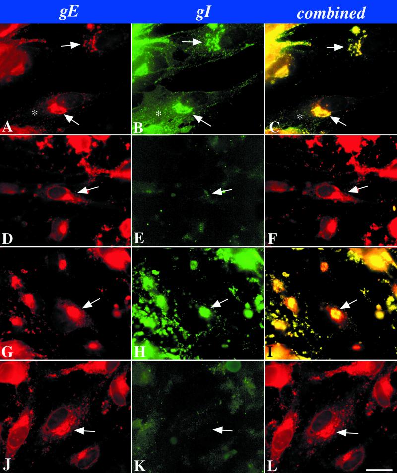
FIG. 2.
Immunocytochemical localization of gI and gE immunoreactivities in cells infected with intact (Ellen) or mutant VZV as shown in Fig. 1. (A to C) Wild-type VZV. (D to F) gIΔ. (G to I) gIΔC. (J to L) gIΔN. The arrows point to the location of the TGN in the infected cells. The asterisk indicates plasma membrane immunostaining. Bar, 10 μm.

