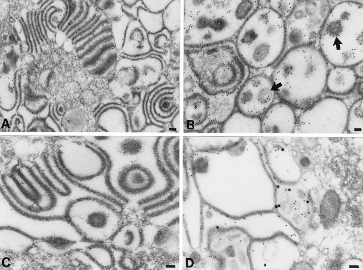
FIG. 6.
The membranes of adherent sacs in the TGN of cells infected with the gIΔC mutant virus display both gE and gI immunoreactivities. (A and B) gE immunoreactivity, demonstrated with 10-nm particles of immunogold in the region of the TGN. (A) Gold particles are concentrated in the membranes of adjacent sacs and are not found in the cytosol. (B) The honeycomb-like structure (see Fig. 5D) found in the region of the TGN of a heavily infected cell contains gE immunoreactivity. Both of the membranes that delimit the chambers of the honeycomb and the virions within the cavities of the chambers are gE immunoreactive. The peripheral distribution of the gE immunoreactivity around the virions (B, arrows) indicates that gE is located in the viral envelope. (C) gI immunoreactivity, demonstrated with 10-nm particles of immunogold in the region of the TGN. Gold particles are concentrated in the membranes of adjacent sacs. The immunoreactivity of gI is less abundant than that of gE (compare with panel A). (D) gE immunoreactivity, demonstrated with 10-nm immunogold particles and gI immunoreactivity demonstrated simultaneously with 20-nm immunogold particles. The gE and gI immunoreactivities are each located in the same membranes lining the chambers of the honeycomb found in the TGN of a highly infected cell. Bars, 150 nm.

