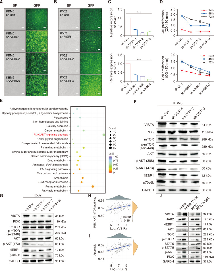
Fig. 7.
VSIR promotes the proliferation of CML cells through regulation of the PI3K/AKT/mTOR pathway ex vivo. (A, B) KBM5 and K562 cells were transfected with FITC-conjugated sh-VSIR vectors. Significant green fluorescence was observed in sh-VSIR-transfected KBM5 cells (bar: 100 μm). (C) The relative mRNA expression of VSIR in sh-VSIR-transfected cells and sh-con cells (p<0.001). (D) CCK-8 results showing that VSIR knockdown inhibited the proliferation of KBM5 (p<0.001) and K562 cells (p<0.05). (E) KEGG pathway analysis showing enrichment of differentially expressed genes (DEGs) between KBM5-sh-con and KBM5-sh-VSIR-3. The enriched KEGG signaling pathways were selected to demonstrate the primary biological actions of the DEGs. For the enrichment results, p<0.05 or FDR<0.05 was considered to indicate a meaningful pathway (an enrichment score of −log10 (P) greater than 1.3). (F, G) Western blots showing that knockdown of VSIR in KBM5 and K562 cells significantly downregulated the PI3K/AKT/mTOR signaling pathway (n=3). (H, I) The correlations between the VSIR gene and the PI3K/AKT/mTOR (p<0.001, ρ=0.36) and apoptosis (p<0.001, ρ=0.38) pathway scores were analyzed with Spearman’s correlation coefficient. (J) Western blots showing that VSIR overexpression in KBM5 and K562 cells significantly increased the activity of the PI3K/AKT/mTOR pathway. Asterisks (*) indicate significance, *** indicates p<0.001.

