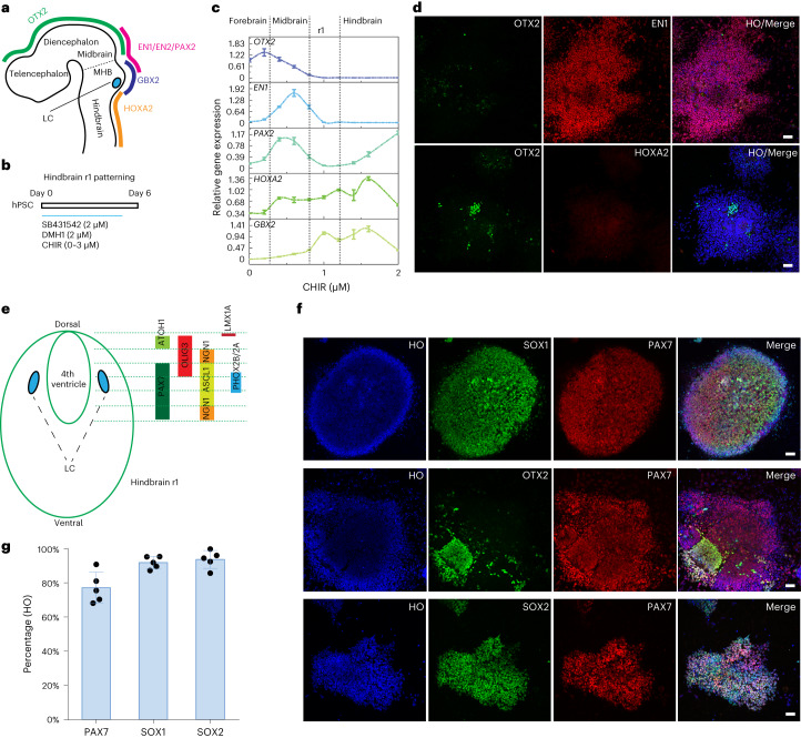
Fig. 1. Specification of dorsal hindbrain (r1) neuroepithelia.
a, Schematic representation of LC location along the embryonic forebrain, midbrain and hindbrain and their corresponding homeodomain transcription factors. b, Experimental design to pattern hindbrain r1 region from hPSCs during the first 6 d of neural induction. c, Expression of forebrain, midbrain and hindbrain genes under a series of CHIR99021 (CHIR) concentrations. Data are shown as mean ± s.e.m. n = 3 biologically independent samples for each condition. d, Immunostaining for OTX2, EN1 and HOXA2 in day 6 cells when treated with 1.0 µM CHIR99021 in the presence of SB431542 and DMH1. Scale bar, 50 µm. e, Schematic representation of the hindbrain r1 domains along the dorsal to ventral subdomains and their corresponding transcription factors. f, Immunostaining for PAX7, SOX1 and SOX2 at day 6 from cells treated with 1.0 µM CHIR99021 in the presence of SB431542 and DMH1. HO, Hoechst. Scale bar, 50 µm. g, Quantification of SOX2-, SOX1- and PAX7-expressing cells at day 6 when treated with 1.0 µM CHIR99021. Data are shown as mean ± s.d. n = 5 biologically independent samples for each condition.

