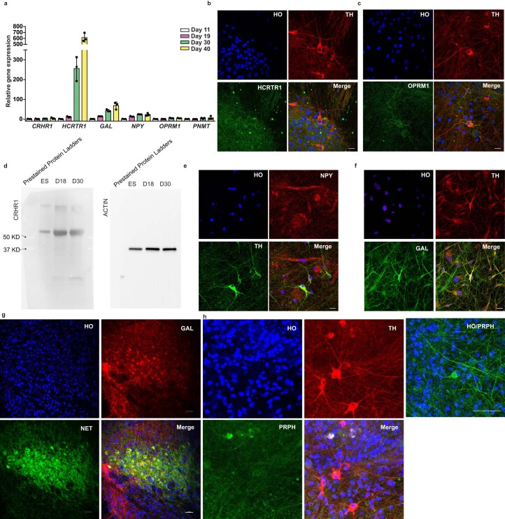
Extended Data Fig. 4. Characterization of hPSC derived NE neuron.
a, qPCR of gene expression following NE neuronal differentiation at day 11, 19, 30 and 40. Data are shown as mean ± s.d. n = 3 biologically independent samples for each condition. b, c, Immunostaining for HCRT1 and OPRM1 in H9 derived NE neurons at day 30. Scale bars, 20 μm. d, The expression of CRHR1 by western blot at Day 0 (ES), Day 18 (D18) and Day 30 (D30) along NE differentiation. e, f, Immunostaining for NPY and GAL in H9 derived NE neurons at day 30. scale bars, 50 μm. g, Immunostaining for GAL and NET in mouse LC to demonstrate the specificity of the GAL antibody. Scale bar, 50 μm. h,Immunostaining for PRPH (Peripherin) in hPSC derived LC-NE neurons at day 30. A positive control to demonstrate the specificity of the peripherin antibody is provided using hPSC derived neurons. Scale bars, 20 μm.

