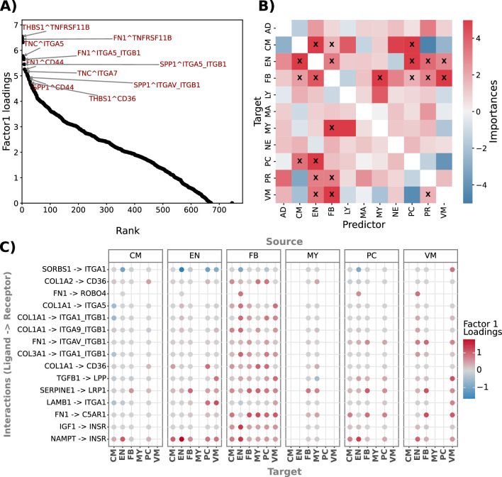
Extended Data Fig. 6. Condition-relevant ligand-receptor interactions and spatially-co-localized cell types in Myocardial infarction.
A) Top 30 interactions (with ‘FN1’, ‘TNC’, ‘THBS1’, and ‘SPP1 as ligands) in Factor 1 identified using NMF on local ligand-receptor metrics in spatially resolved 10X Visium heart samples. B) Importances (Median t-values clipped at 5 and -5; n = 28) from a spatially-weighted model predicting all possible cell type interactions from Myocardial infarction 10X Visium slides. Cell type interactions with Median t-value > 1.645 and R2 > 5% are marked with X. C) Multi-view Factorization feature (interaction) loadings following decomposition of ligand-receptor scores inferred from dissociated single-nucleus myocardial infarction data. The top 15 interactions with the highest interaction loadings are shown across all cell-type pairs. Abbreviations used include AD for Adipocytes, CM for Cardiomyocytes, EN for Endothelial cells, FB for Fibroblasts, PC for Pericytes, PR for Proliferating cells, VM for Vascular smooth muscle cells, NE for Neuronal cells, MY for Myeloid cells, MA for Mast cells, and LY for Lymphoid cells. Source numerical data are available in source data.

