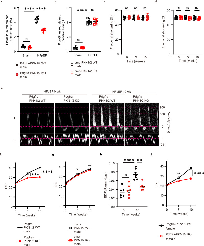
Fig. 8. PKN 1/2 deletion in resident cardiac fibroblasts, not in cardiomyocytes, suppressed cardiac fibrosis in HFpEF.
Fibrotic changes in left ventricles assessed using Picrosirius red staining at 10 weeks after combination exposure to HFD and L-NAME in Pdgfra-PKN1/2 WT and KO mice (a) and in cardiomyocyte-specific PKN1/2 (cmc-PKN1/2) WT and KO mice (b) (n = 6, biological replicates per group). ns, not significant; ****p < 0.0001. Fraction shortening examined using echocardiography at 0, 5, and 10 weeks after combination exposure to HFD and L-NAME in Pdgfra-PKN1/2 WT and KO mice (c) (n = 10, biological replicates per group) and in cmc-PKN1/2 WT and KO mice (d) (n = 6, biological replicates per group). ns, not significant. e Representative early diastolic mitral inflow velocity (top) and annular velocity (bottom). Mitral E and E’ waves (E/E’) ratio at 0, 5, and 10 weeks after combination exposure to HFD and L-NAME in Pdgfra-PKN1/2 WT and KO male mice (f) (n = 10, biological replicates per group), and in cmc-PKN1/2 mice WT and KO male mice (g) (n = 6, biological replicates per group). ns, not significant; ***p = 0.0006; ****p < 0.0001. h End-diastolic pressure-volume relationship (EDPVR) obtained by cardiac catheterization at 0 and 10 weeks after combination exposure to HFD and L-NAME in Pdgfra-PKN1/2 WT and KO mice (n = 6, biological replicates per group). ns, not significant; **p = 0.0035; ****p < 0.0001. i E/E’ at 0, 5, and 10 weeks after combination exposure to HFD and L-NAME in Pdgfra-PKN1/2 WT and KO female mice (n = 6, biological replicates per group). ns, not significant; ****p < 0.0001. Data are presented as the mean ± SEM and analyzed using two-way ANOVA followed by Tukey’s post hoc test (a–d, h), or with two-way repeated measures ANOVA followed by Bonferroni post hoc test (f, g, i). The data represent three independent experiments with similar results. Source data are provided as a Source Data file.

