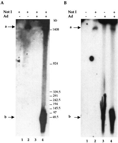
FIG. 4.
Analysis of rep-cap amplified DNA molecules by pulsed-field gel electrophoresis. Samples for pulsed-field gel electrophoresis were prepared from noninfected or adenovirus-infected HeRC32 cells (MOI, 50) as described in Materials and Methods and were analyzed using a rep probe (1.4 kb). Where indicated, DNA was digested with NotI, which does not cut in the rep-cap genome. (A) Lanes 1 and 2, noninfected HeLa and HeRC32 cells, respectively; lanes 3 and 4, adenovirus-infected (48 h) HeLa and HeRC32 cells, respectively. (B) Lanes 1 and 2, noninfected HeRC32 cells; lanes 3 and 4, adenovirus-infected (48 h) HeRC32 cells. The two arrows indicate the positions of the integrated (a) and extrachromosomal (b) rep-cap fragments.

