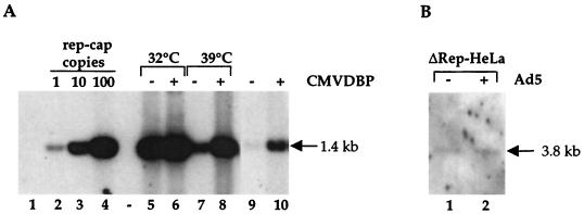
FIG. 6.
(A) Effect of the adenovirus DBP on rep-cap amplification. HeRC32 cells were infected with Ad.ts125 (MOI, 50) at the indicated temperature, and total DNA was analyzed by Southern blotting using a rep probe (1.4 kb) as described in the legend to Fig. 1. Where indicated, the CMVDBP plasmid (10 μg) was transfected into 4 × 10⁶ HeRC32 cells using Exgen (EuroMedex), either alone or 6 h prior to adenovirus infection. In this case, the transfection was done at 37°C and the cells were switched to the indicated temperature immediately after adenovirus infection. Lane 1, DNA from noninfected HeLa cells; lanes 2, 3, and 4, standard rep-cap genome copies; lane 5, DNA from HeRC32 cells infected with Ad.ts125 at 32°C; lane 6, DNA from HeRC32 cells transfected with CMVDBP and infected with Ad.ts125 at 32°C; lane 7, DNA from HeRC32 cells infected with Ad.ts125 at 39°C; lane 8, DNA from HeRC32 cells transfected with CMVDBP and infected with Ad.ts125 at 39°C; lane 9, DNA from noninfected HeRC32 cells; lane 10, DNA from HeRC32 cells transfected with the CMVDBP plasmid. (B) Analysis of rep-cap amplification in ΔRep-HeLa cells. Total DNA was extracted from uninfected (lane 1) and adenovirus-infected (lane 2) ΔRep-HeLa cells, digested with PstI, and analyzed on a Southern blot as previously indicated. Since the deletion in the rep sequence removes one PstI site, the size of the expected band is 3.8 kb.

