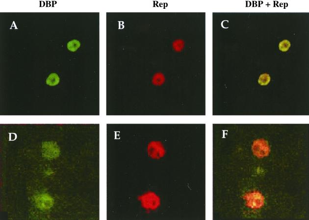
FIG. 8.
Detection of Rep and DBP proteins following transfection of the CMVDBP plasmid into HeRC32 cells. A total of 6 × 10⁴ HeRC32 cells grown on glass slides were transfected with 0.4 μg of the CMVDBP plasmid. Forty-eight hours later, the cells were fixed and analyzed by immunofluorescence using an anti-DBP (26) and an anti-Rep 68/40 (A, B, and C) or anti-Rep 78/52 (D, E, and F) antibody (39). Cells were photographed with either a fluorescein (A and D) or a rhodamine (B and E) filter. In panels C and F, the two images are superimposed. Magnification, ×1,000.

