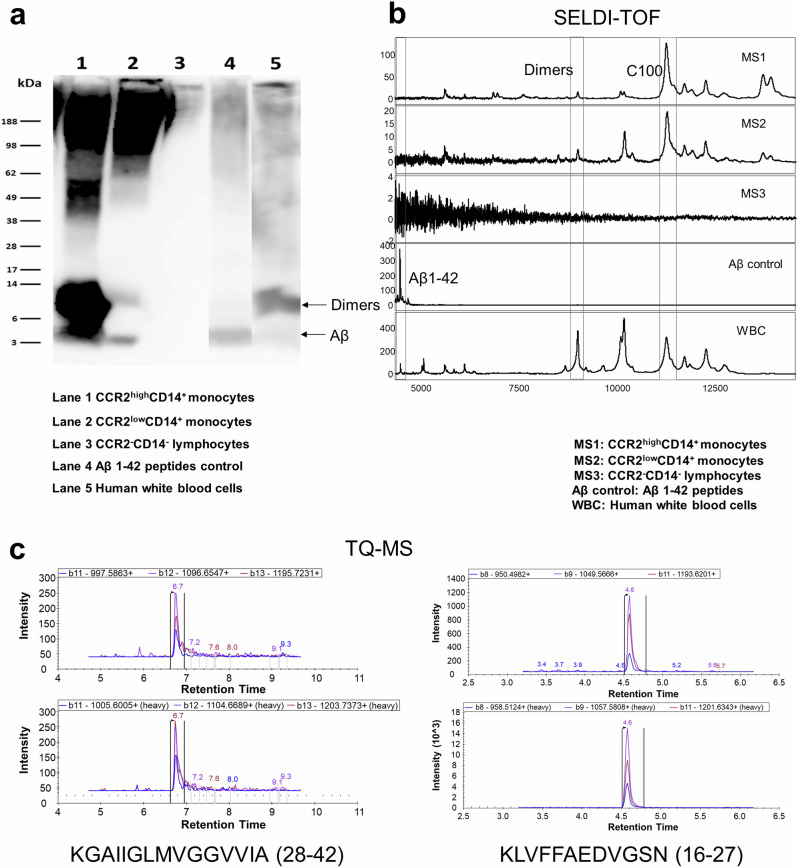
Fig. 3. Confirmation of Aβ1-42 presence on Aβ-binding monocytes in blood.
PBMCs from CU individuals were FACS sorted based on CCR2 and CD14 expression. a Western blots (n = 2) showed substantial Aβ1-42 on CCR2++CD14+ monocytes, smaller amounts on CCR2+CD14+ monocytes, and no signal on CCR2-CD14- lymphocytes. b SELDI-TOF mass spectrometry (n = 2) confirmed diverse Aβ1-42 species, including dimers, in CCR2++CD14+ and CCR2+CD14+ monocytes, but not in CCR2−CD14− lymphocytes. c TQ-MS (n = 3) identified Aβ-specific fragments (28–42) on various cell types, including CCR2++CD14+ and CCR2+CD14+ monocytes, and CCR2−CD14− lymphocytes, with fragment (16–27) also detected abundantly.

