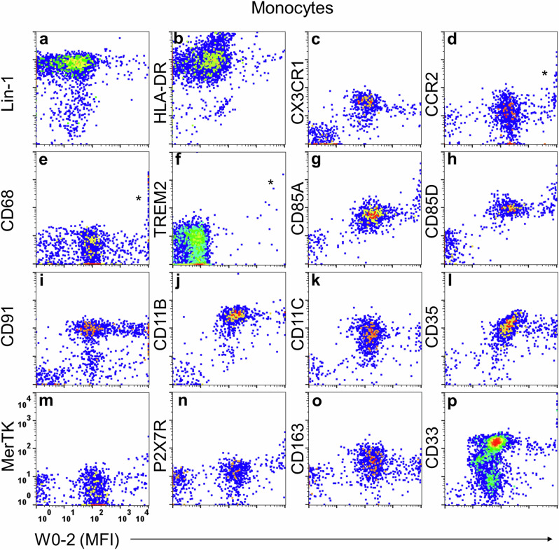
Fig. 4. Characterization of Aβ-binding monocytes.
Peripheral Aβ-binding monocytes (n ≥ 3, with a total of 22 blood samples analyzed) were analyzed using four-color immunophenotypic analysis. Aβ-binding monocytes exhibited a LIN-1+ and HLA-DR+ (MHC-II) phenotype (a, b), indicating differentiation from dendritic cells, and expressed CX3CR1 and CCR2 chemotactic receptors (c, d), suggesting strong migratory capabilities. They also showed CD68+ macrophage-like (e) and TREM2+ microglia-like phenotypes (f). Aβ-binding monocytes expressed Aβ-binding receptors (CD85A, CD85D, CD91) (g–i), complement receptors (CD11b, CD11c, CD35) (j–l), and phagocytosis-associated receptors (MerTK, P2X7R, CD163, CD33) (m–p). It is worth noting that in the four-color panel for TREM2 (f), what matches TREM2, CD14, and CD16 was Qdot525 conjugated W0-2, unlike in the other panels where W0-2 was used in conjunction with a secondary antibody.

