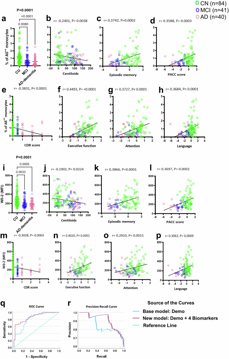
Fig. 5. Peripheral monocyte surface Aβ as a potential biomarker for AD.
Quantifying Aβ on peripheral monocytes may serve as a non-invasive biomarker for AD. Our study included 150 participants (78 CU, 36 MCI, 36 AD-dementia). a–p A decrease in the percentage of Aβ++ monocytes in MCI and AD-dementia compared to CU. Fluorescence intensity also showed a decrease in MCI and AD-dementia. A negative correlation was observed between the percentage of Aβ++ monocytes and brain Aβ-PET burden. Similarly, fluorescence intensity is weakly correlated with the Aβ-PET burden. Correlations between the percentage of Aβ++ monocytes, fluorescence intensity, and cognitive function were also observed, showing moderate correlations. The datasets were not normally distributed, and P-values were determined using the Kruskal-Wallis test, followed by Dunn’s multiple comparisons test (solid zig-zag line). The column dot plots represent mean ± standard deviation. Correlation coefficients (r) and P-values were calculated using Spearman correlation analysis. q, r A multivariate model revealed the potential of monocyte surface Aβ in predicting brain Aβ-PET burden, with a panel of %Aβ++ monocytes, %Aβ+ NK cells, %Aβ++ classical monocytes, and %classic monocytes performing best (AUC = 0.871). The new panel significantly improved AD discrimination, with a sensitivity of 0.65 and specificity of 0.976.

