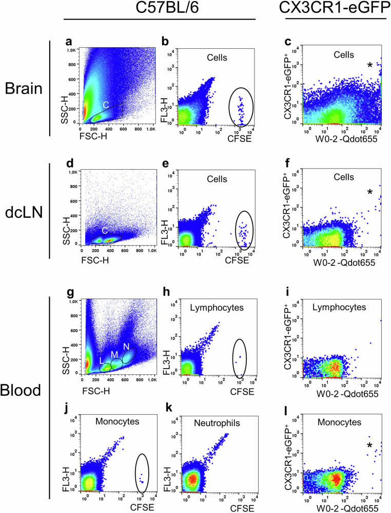
Fig. 7. Detection of Adoptive PBMCs in Blood and Deep Cervical Lymph Nodes.
PBMCs were sourced from donor C57BL/6 mice and labeled with CFSE (n = 13) or from CX3CR1-eGFP mice without CFSE labeling (n = 5). Both sets of PBMCs were injected into the lateral ventricles of APP/PS1 mice between 15 and 80 weeks of age. Two days later, recipients’ brains, deep cervical lymph nodes (dcLN), and blood were examined using fluorescent flow cytometry. a, d, and g display typical flow cytometry light scatter dot plots of the brain, dcLN, and blood, respectively. The presence of donors’ CFSE+ cells in the recipients’ brains confirmed successful delivery (circled in b). Likewise, CFSE+ cells in recipients’ dcLN (circled in e) and blood (circled in h, j) validated peripheral migration. No CFSE+ cells were found in the recipients’ blood neutrophil gating (k). Notably, the donors’ CX3CR1-eGFP+ monocytes were identified as carrying substantial amounts of Aβ in the recipients’ brain (asterisked in c), dcLN (asterisked in f) and blood (asterisked in l). A few CX3CR1-eGFP+ monocytes were found in the recipients’ blood lymphocyte gating (i) due to an overlap between lymphocytes and monocytes. This flow cytometry analysis demonstrates the migration and Aβ-carrying capabilities of adoptive PBMCs in different tissues. C: Cells; L: Lymphocytes; M: Monocytes; and N: Neutrophils.

