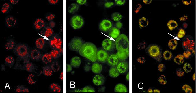
FIG. 5.
Immunofluorescence double colocalization of LANA1 and LANA2 in KSHV-infected BCBL-1 cells. (A) LANA1 protein (red) in a coarsely speckled nuclear distribution. (B) Diffuse, finely speckled nuclear pattern of LANA2 protein (green). (C) Double filter; colocalization of LANA1 and LANA2. (A, B, C) Although some subnuclear regions show the distinct dispersal of the two proteins exclusive of each other, yellow nuclear staining is also evident in other areas, possibly representing colocalization of a subfraction of LANA1 and LANA2. Cells undergoing mitosis (arrow) appear to express only LANA1 exclusive of LANA2 (C) (magnification, ×100; Texas red and fluorescein isothiocyanate).

