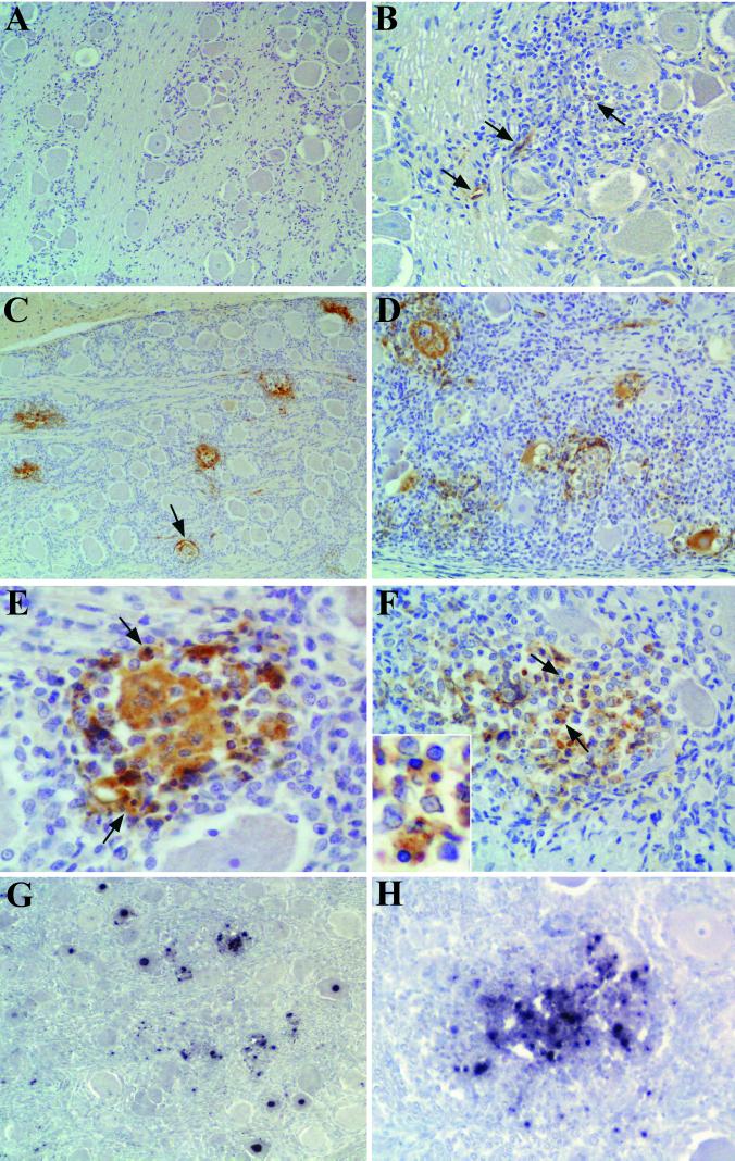
FIG. 2.
Detection of PRV antigen (A to F) by IHC and of PRV DNA (G and H) by ISH in the TG of uninfected and PRV-infected pigs. (A) TG section from the uninfected pig showing no positive immunoreaction. (B) Viral antigen (brown stain) was detected in the cytoplasm of a small number of infiltrating immune cells (arrows) at 24 hpi. (C) At 48 hpi, PRV antigen was present in scattered neurons, in perineuronal inflammatory cells, and in satellite cells (arrow). (D) Multiple foci of inflammation containing PRV antigen-positive neurons and inflammatory cells at 72 hpi. (E) A PRV-infected neuron in the process of being eliminated by immune cells at 48 hpi. Note the presence of viral antigen in inflammatory cells, some of them showing apoptotic morphology (arrows). (F) At 72 hpi, an infected neuron has been degraded and viral antigen is present in many inflammatory cells, some of them exhibiting clearly an apoptotic appearance (arrows and inset). (G) PRV DNA (black stain) was detected in the nuclei of neurons and in infiltrating inflammatory cells at 48 hpi. (H) Focus of neuronophagia containing numerous PRV-infected inflammatory cells at 72 hpi. Original magnifications, ×100 (A, C, D, and G), ×200 (B and H), and ×400 (E and F).

