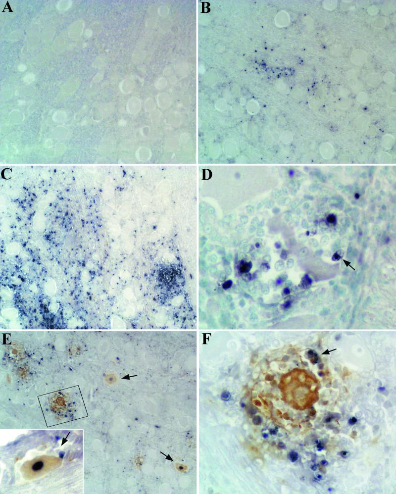
FIG. 3.
Detection of apoptotic cells (A to D) by the in situ TUNEL method and colocalization of PRV antigen and apoptotic cells (E and F) by double labeling in the TG. (A) No apoptotic cells were detected in the TG at 12 hpi. (B) A few apoptotic inflammatory cells (black stain) were observed among infiltrating inflammatory cells at 24 hpi. (C) The maximum number of apoptotic cells was observed at 72 hpi. Note the absence of staining in neurons. (D) Apoptotic cells surrounding a neuron at 72 hpi. An apoptotic body seems to be phagocytosed by a macrophage (arrow). (E) PRV antigen (brown stain) and apoptotic cells (black stain) were distributed in the same regions of the TG. Three TUNEL-positive neurons (arrows and inset) were detected at 48 hpi. Note the presence of immune cells (arrow) in direct apposition to the IHC-positive–TUNEL-positive neuron shown in the inset. (F) Detail of boxed area in panel E under higher magnification. PRV-infected neurons were usually TUNEL negative. A double-labeled apoptotic cell can be identified (arrow). Original magnifications, ×100 (A, B, C, and E) and ×400 (D, F, and E [inset]).

