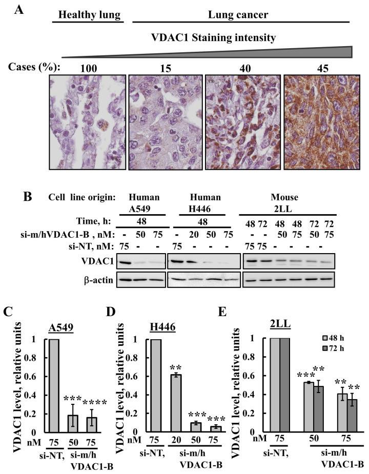
Figure 1.
VDAC1 is overexpressed in lung cancer and VDAC1 expression is silenced in lung cancer cell lines using si-m/hVDAC1-B. (A) IHC staining with anti-VDAC1 antibodies was performed on lung tissue microarray slides (US Biomax). Representative images from sections of the NSCLC tissues (n = 31) and healthy pulmonary tissues (n = 10) with the percentage of sections stained at the intensity indicated by the scale above. Sections of the tissues were observed under an Olympus microscope, and images were taken at 600× magnification. (B) Lung cancer cell lines A549, H446 and 2LL were transfected with the indicated concentration of si-m/hVDAC1-B using silenFect transfection agent, as described in the Methods sections. Cells were harvested 48 or 72 h post transfection and subjected to immunoblotting using anti-VDAC1 antibodies. (C–E) Quantification of the VDAC1 expression levels in the three cell lines. Results present the mean ± SD (n = 3), ** p ≤ 0.01, *** p ≤ 0.001, **** p ≤ 0.0001. Original western blots are presented in File S1.

