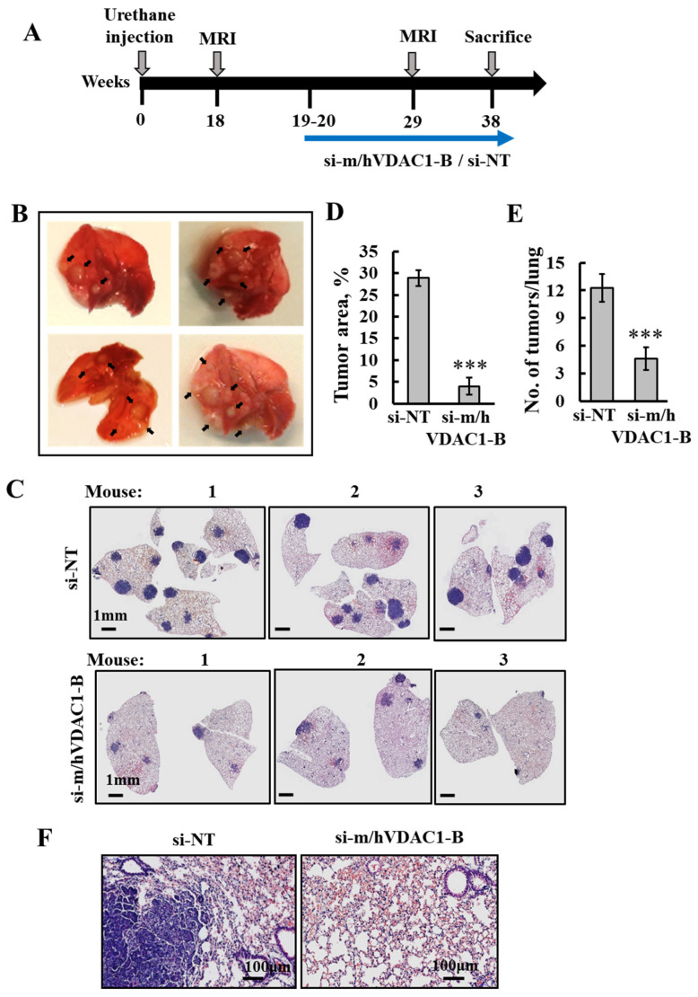
Figure 2.
PLGA-PEI-si-m/hVDAC1-B treatment of urethane-induced lung cancer reduced tumor size and number. (A) Schematic illustration of the study design: A/J mice, 7–8 weeks old, were injected i.p. with urethane (1 mg/gr body weight), and tumor development and treatment are indicated. (B) Photo of lungs from mice sacrificed at week 19, after exposure to urethane, with the arrows pointing to tumors. Following tracking of tumor development with MRI at week 18, treatment with PLGA-PEI-si-m/hVDAC1 or PLGA-PEI-si-NT (200 nM blood concentration) was started at week 19, and performed three times a week until the mice were sacrificed at week 38. (C,F) Representative H&E staining of paraffin-embedded, formaldehyde-fixed sections of the lungs after treatment with PLGA-PEI-si-m/hVDAC1-B or PLGA-PEI-si-NT. (D,E) Quantitative analysis of tumor area presented as a % of the total section area (PLGA-PEI-si-NT, n = 7, PLGA-PEI-si-m/hVDAC1, n = 8) (D), and the number of tumors (PLGA-PEI-si-NT n = 4 and PLGA-PEI-si-m/hVDAC1-B, n = 4) (E). Results reflect the mean ± SEM, *** p ≤ 0.001. (F) Enlargement of a selected area from the lung shown in C.

