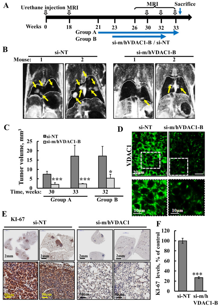
Figure 3.
PLGA-PEI-si-m/hVDAC1-B treatment of urethane-induced lung cancer decreased VDAC1 expression level and inhibited tumor growth. (A) Schematic illustration of the study design: A/J mice, 7–8 weeks old, were injected i.p. with urethane (1 mg/gr body weight). Treatment with PLGA-PEI-si-m/hVDAC1 or PLGA-PEI-si-NT (350 nM) was started at week 21 (group A) or at week 23 (group B) and continued three times a week until the mice were sacrificed. MRI imaging was performed at weeks 18, 30, 32, and 33. Mice were sacrificed at week 33 (group A) and week 32 (group B). (B) Representative MRI images at week 33 (group A) of PLGA-PEI-si-NT- and PLGA-PEI-si-m/hVDAC1-B-treated mice. The arrows point to tumors. (C) Quantification of tumor volume from MRI images of group A at week 30 (PLGA-PEI-si-NT, n = 6, PLGA-PEI-si-m/hVDAC1-B, n = 7) and week 33 (PLGA-PEI-si-NT, n = 5, PLGA-PEI-si-m/hVDAC1-B, n = 5) and for group B at week 32 (PLGA-PEI-si-NT, n = 3, PLGA-PEI-si-m/hVDAC1-B, n = 4). (D) IF staining of VDAC1 in lung sections from PLGA-PEI-si-m/hVDAC1-B and PLGA-PEI-si-NT-treated mice. The squares point to the area enlarged below. (E,F) IHC staining for KI-67 using specific antibodies of PLGA-PEI-si-NT- and PLGA-PEI-si-m/hVDAC1-B-treated mice (E). For the sections, the lungs with tumors are shown with enlarged area below. KI-67 staining intensity was analyzed quantitatively (n = 7) (F). Results present the mean ± SEM, * p ≤ 0.05, *** p ≤ 0.001.

