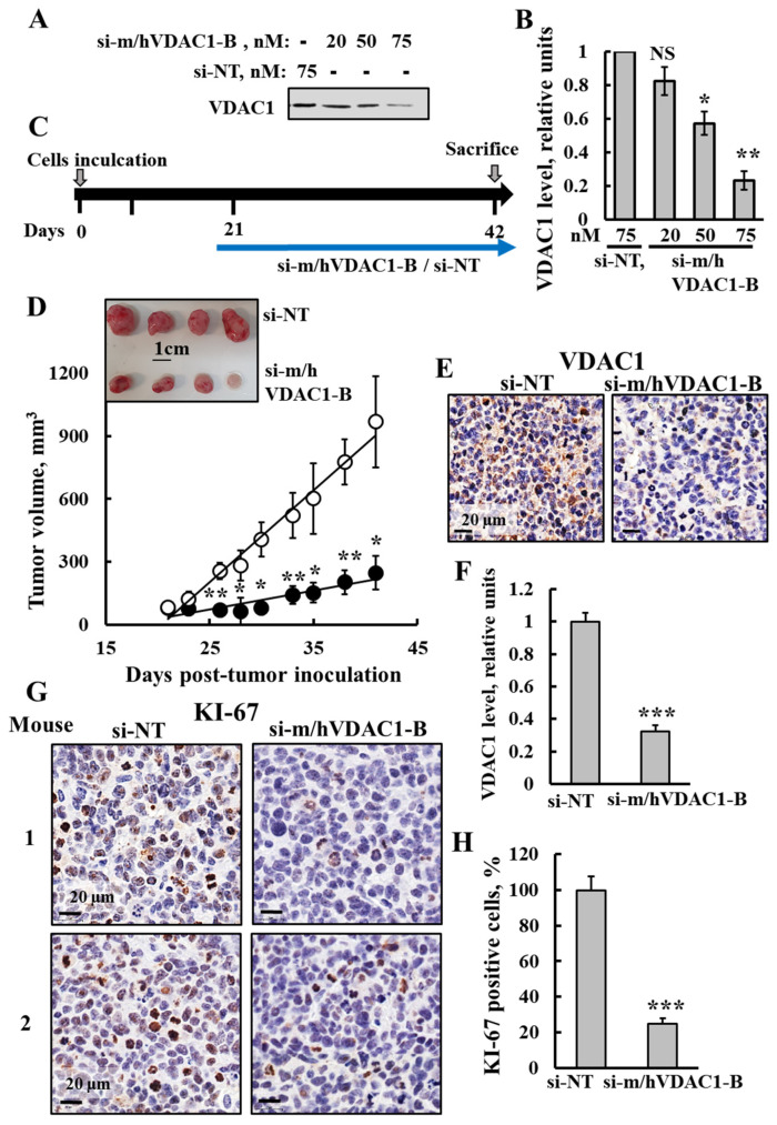
Figure 7.
si-m/hVDAC1-B tumor treatment inhibited tumor growth, reduced VDAC1 expression and cell proliferation in an SCLC xenograft mice model. (A,B) SCLC H69 cells were transfected with the indicated concentration of si-NT or si-m/hVDAC1-B using silenFect transfection agent, as described in the Methods sections. Cells were harvested 48 h post transfection and subjected to immunoblotting using anti-VDAC1 antibodies (A) and VDAC1 expression level quantification (B). (C) Schematic illustration of the study design: H69 cells (2 × 10⁶ cells/mouse) were inoculated into Athymic (C57BL/6JOlaHsd) 6-week-old male mice. When the tumor volume was 50–80 mm3 (at day 21), the mice were sub-divided into two groups: si-m/hVDAC1-B (200 nM) and si-NT (200 nM) -treated xenografts, injected three times a week and the mice were scarified at day 42. (D) Tumor size was measured (using a digital caliper) and tumor volume was calculated with the average presented as the mean ± SEM.; si-m/hVDAC1-B (●, n = 5) and si-NT (o, n = 4). A photograph of the tumors excised from sacrificed mice is also shown (inset). (E,F) Representative IHC-stained tumor sections of si-m/hVDAC1-B- and si-NT-treated mice using specific antibodies against VDAC1 (E) and their quantified staining intensity (F). (G,H) Representative IHC-stained tumor sections of si-m/hVDAC1-B- or si-NT-treated tumors using specific antibodies against KI-67 (G). Quantitative analysis of KI-67 positive cells (H). Results reflect the mean ± SEM (n = 4–5), * p ≤ 0.05, ** p ≤ 0.01, *** p ≤ 0.001. Original western blots are presented in File S1.

