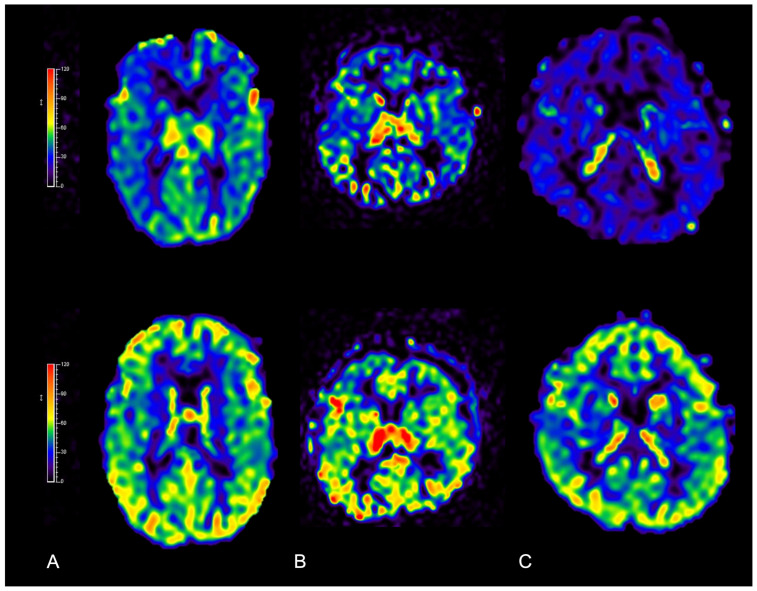
Figure 14.
Qualitative increase in cerebral blood flow demonstrated with ASL perfusion MRI before (upper row) and 6 months after surgery (bottom row) in three patients diagnosed with nonsyndromic isolated CRS: (A) scaphocephaly; (B) anterior plagiocephaly; and (C) trigonocephaly. Threshold color scale bars are shown on the left.

