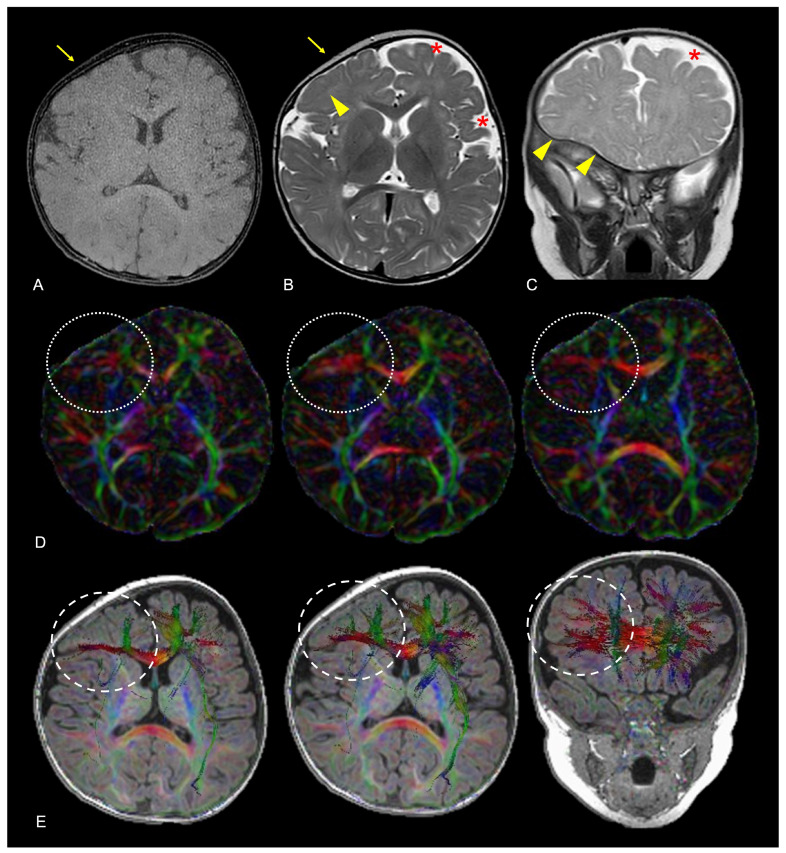
Figure 15.
Axial black-bone MR image (A), axial TSE T2W image (B) and coronal TSE T2W image (C) of a 9-month-old female patient diagnosed with anterior plagiocephaly due to premature fusion of the right coronal suture (yellow arrows); aberrant adaptation of the right frontal lobe nervous tissue is indirectly demonstrated by the anomalous orientation of the frontal and frontal-insular sulci both on the axial and coronal planes (yellow arrowheads) compared to the left side, coupled to the asymmetric representation of adjacent CSF spaces (red asterisks). Axial FA maps (D) and color-coded representation of diffusion tensors on axial and coronal reconstructions superimposed on 3D-T1W images (E) confirmed altered fractional anisotropy (white dotted lines) and aberrant white matter fiber orientation (white dashed lines) due to white matter structural adaptive changes in the corresponding area. Color legend: red for left–right; blue for superior–inferior; and green for anterior–posterior.

