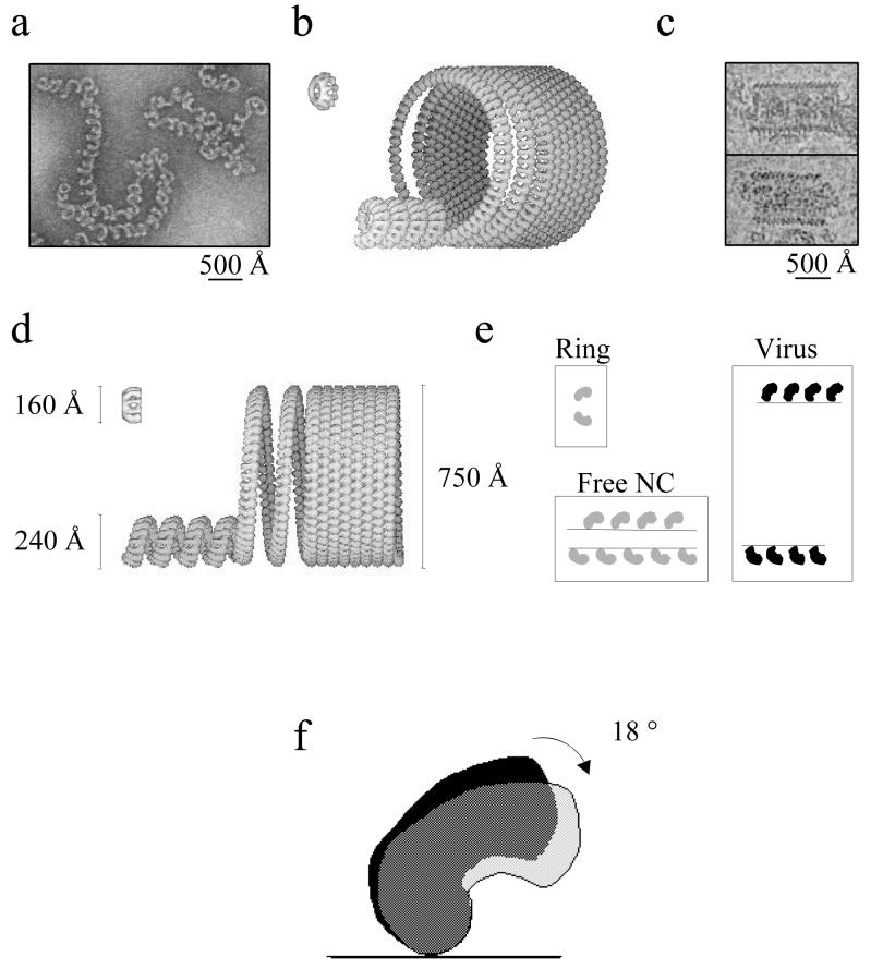
FIG. 6.
Models of free nucleocapsid and nucleocapsid inside virus. Negatively stained images of viral nucleocapsids isolated with CsCl gradient centrifugation (a) and cryo-EM images of rabies virus skeletons that were present in an untreated virus preparation (c) were used to determine the diameter of the two helical structures shown in panels b and d. (b) Tilted view of the recombinant rings, the free nucleocapsid, and nucleocapsid inside virus; (d) side views with an indication of the diameters. (e) Tilt angles between N and the symmetry axis of the rings or the helical axes in the two types of nucleocapsids. The difference of 18° between the tilt angles of N in rings or free nucleocapsids and N in virus is also shown (f).

