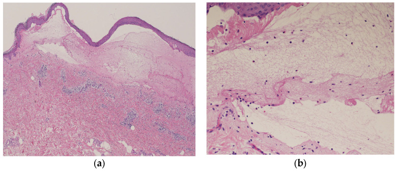
Figure 5.
Histopathological examination of the blistered area reveals subepidermal blisters (hematoxylin and eosin [HE] staining; ×40) (a); eosinophils and lymphocytes were infiltrating around the small blisters, along with marked infiltration of inflammatory cells in the upper dermis (HE staining; ×200) (b).

