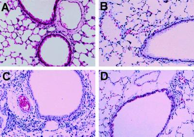
FIG. 4.
Histology of the lung after the final challenge (hematoxylin-eosin staining). In the acute phase group (C), infiltration of mononuclear cells and eosinophils was observed in the submucosa. In the recovery phase group (D), little infiltration of mononuclear cells was observed in the submucosa and no eosinophils were detected in the lung. No inflammatory cell infiltration was observed in the control (A) and OA (B) groups. Magnification, ×100.

