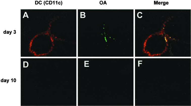
FIG. 5.
Double coloring of the lung 2 h after OA inhalation on day 3 (A to C) or day 10 (D to F) (DCs are stained red; FITC-labeled OA is green). In the acute phase group (day 3), DCs stained with the MAb of CD11c migrated to the bronchial epithelium (A), and OA was observed at the same site of the lung (B). Merging of the two images shows OA-capturing APCs (in yellow) on the bronchial epithelium (C). In the recovery phase group (day 10), neither DCs nor OA was detected anywhere in the lung (D and E). Magnification, ×50.

