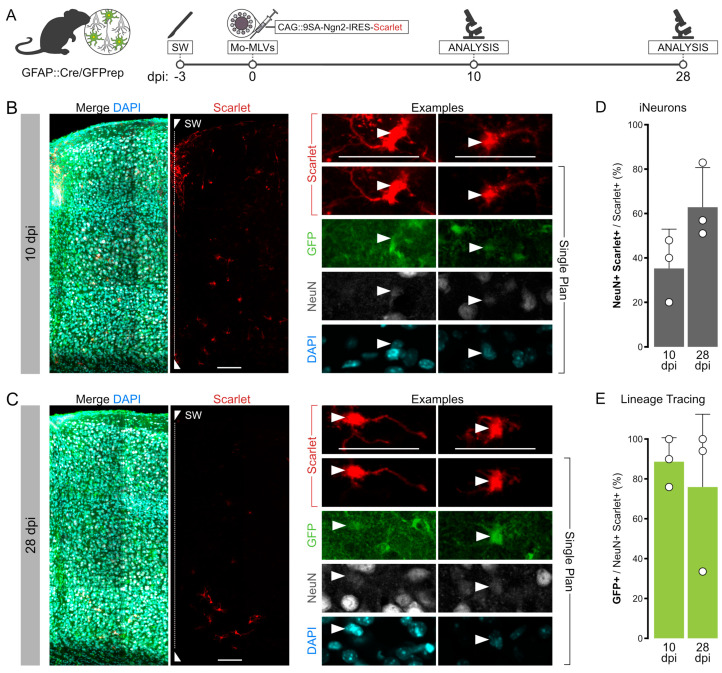
Figure 4.
Genetic lineage tracing of endogenous astrocytes during Mo-MLVs transduction. (A) Schematic of the surgical procedures used to lineage trace endogenous astrocytes in SW-injured GFAP::Cre/GFPrep mice after the injection of Mo-MLVs. (B,C) Confocal pictures of a 15 μm stack of SW-injured motor cortex of GFAP::Cre/GFPrep mice at 10 (B) or 28 (C) days after the injection of Mo-MLVs-CAG::9SA-Ngn2-IRES-mScarlet. Scalebar 50 μm. Single optical plane magnifications of a representative group of cells are reported in inserts I-IV. Mo-MLVs-transduced cells (red) were double-stained for the lineage tracing reporter GFP (green) and neuronal marker NeuN (white). Scalebars: 50 μm. (D,E) Quantification and lineage tracing analysis of the NeuN+/mScarlet+ cells. Plotted data represent the mean ± SD of three biological and technical replicates.

