Figure 3.
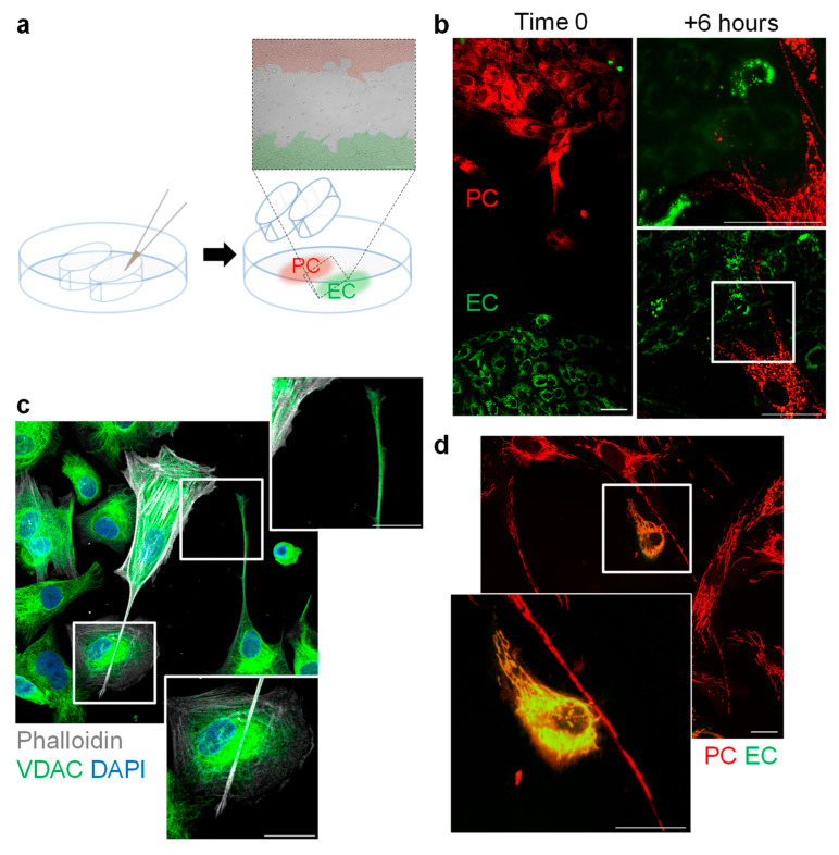
Transfer of mitochondria from pericytes to endothelial cells in vitro. (a) Scheme showing the migration chamber used to study the early contact of MitoTracker Red-labelled pericytes (PC) with MitoTracker Green-loaded endothelial cells (EC) for up to 6 h. (b) Labelled mitochondria in pericyte TNTs during first contact with endothelial cells in the presence of PDGF-BB (1 ng/mL) for six hours. Comparable results were made using five independent cell batches; bar = 20 µm. (c) Presence of the VDAC in pericyte TNTs. Comparable observations were made using four additional cell batches; bar = 20 µm. (d) Transfer of pericyte-derived mitochondria (red) to endothelial cells (green) in cells cocultured for 6 h in the presence of PDGF-BB (1 ng/mL); bar = 50 µm in overview, 10 µm in magnification. Comparable observations were made using six additional cell batches.

