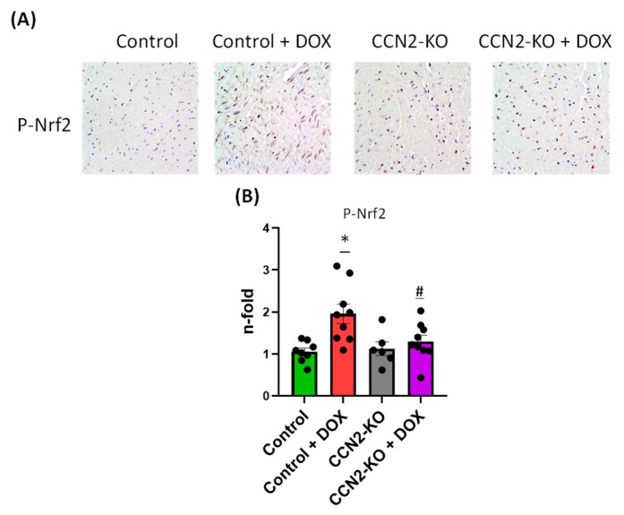
Figure 5.
Immunohistochemistry (IHC) detection of NRF2 protein activation in the left ventricular tissue of mouse hearts. (A) Representative IHC images showing phospho-NRF2 staining. (B) Quantification of positive staining for phospho-NRF2 (p-NRF2) levels. Five days of DOX administration resulted in significantly elevated phospho-NRF2 levels in the Control group mice. In contrast, the CCN2-KO + DOX group did not show significant changes in protein activation compared to their corresponding CCN2-KO control group. * p < 0.05 vs. Control; # p < 0.05 vs. Control + DOX. n = 6–10.

