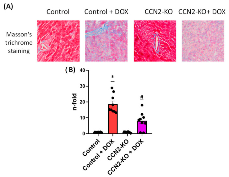
Figure 7.
Cardiac fibrosis in mice assessed using Masson’s trichrome staining of left ventricular (LV) sections. (A) Representative images of the stained sections. (B) Quantitative analysis revealed a significant increase in fibrosis in both DOX-treated groups compared to the untreated groups. However, the Control + DOX group exhibited a greater extent of fibrosis than the CCN2-KO + DOX group. * p < 0.05 vs. Control; # p < 0.05 vs. Control + DOX. n = 6–10.

