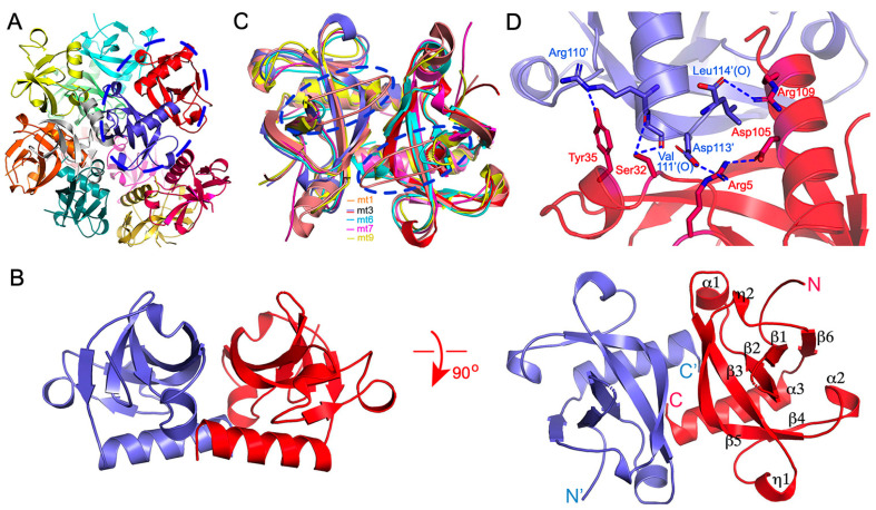
Figure 1.
Overall structure of MazF-mt3. (A) The MazF-mt3 assembly in the asymmetric unit in the ribbon rendition. The 12 monomers are color coded, forming 6 dimers. The dimer for the close-up view in (B) is circled. (B) The representative MazF-mt3 dimer in two orthogonal views. The two subunits are colored red and slate, respectively, The N- and C-termini are indicated, and the secondary structure elements of MazF-mt3 are labeled. (C) Structural comparison between MazF-mt3 (red and slate) and other toxin members in the M. tb MazF family, which include MazF-mt1 (PDB 6KYS, orange), mt6 (PDB 5CCA, cyan), mt7 (PDB 5XE2, pink), and mt9 (PDB 5WYG, yellow). The loop regions are circled by the broken ovals. (D) Close-up view of the dimer interface at the C-termini. The side chains of the key interfacial residues are shown as sticks and labeled. The hydrogen-bonding and salt-bridge interactions are indicated by the blue dashed lines.

