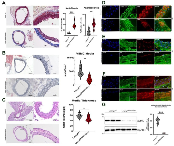
Figure 2.
Histological and Immunofluorescent characterization of thoracic aorta pathology in LmnaG609G/G609G mice compared to wildtype C57B/6 mice. (M = media; A = adventitia, I = intima). Histopathological and immunofluorescent staining at 10× and 40× magnification using the Keyence BZ-X810 microscope and an Axio Imager D2: (A) Media and adventitia fibrosis was assessed using Masson’s Trichrome staining. Significant increase was detected in the adventitia and inside the media in between the elastin fibers (n = 6; *** p < 0.0005; scale bar = 200 µm (10×)/50 µm (40×)). (B) Oil Red O staining counterstained with Hematoxylin to visualize nuclei in aortic media. Media cellularity was found significantly reduced in LmnaG609G/G609G, no lipid staining was observed except in periaortic adipose tissue surrounding the aorta (n = 4; ** p < 0.005; scale bar = 200 µm (10×)/50 µm (40×); arrow = nuclei). (C) Media thickness of thoracic aorta was compared between Lmna+/+ and LmnaG609G/G609G mice. LmnaG609G/G609G mice had a reduced media thickness compared to wildtype animals (n = 6; * p < 0.05; scale bar = 200 µm (10×)/50 µm (40×)). (D) LaminA/C (green), αSMA [35], and 4′,6-diamidino-2-phenylindole (DAPI) staining in aorta; αSMA staining together with DAPI indicates loss of VSMC in LmnaG609G/G609G media. (n = 3; scale bar = 20 µm). Zoom-box was added to show Lamin nuclear rim staining. (E) p16 (green), Vimentin [35], and DAPI (blue) staining; p16 indicating senescence was predominantly found in the intima and media of LmnaG609G/G609G mice. Vimentin signal was lost in media of LmnaG609G/G609G mice but was detected equally in intima and adventitia of both wildtype and mutant mice (scale bar = 20 µm). Zoom-box was added to show specificity of p16 staining at the intima. White arrows indicate location of p16 staining. (F) Serpine-1 (PAI-1) (green), IL-6 [35], and DAPI (blue) staining. PAI-1 signal visible mostly in adventitial region and epidermal layer of the intima. PAI-1 signal was increased in endothelial cells of the aortic intima of LmnaG609G/G609G mice (scale bar = 20 µm). White arrows indicate PAI-1 staining location. Zoom-box was added to show staining specificity at the intima. (G) Western blot detection of αSMA in LmnaG609G/G609G and Lmna+/+ mice showing significantly reduced levels of this protein in mutant aortas ** p < 0.0005).

