Figure 4.
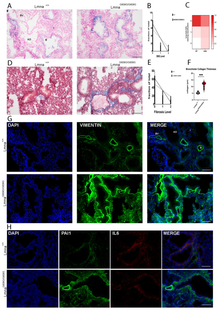
Histological and Immunofluorescent characterization of lung pathology in LmnaG609G/G609G mice compared to wildtype C57B/6 mice. Histopathological and immunofluorescent staining at 10× and 40× magnification using the Keyence BZ-X810 microscope and an Axio Imager D2 (B = bronchiole, AD = alveolar duct; I = Interstitium; BV = blood vessel): (A) Increased β-Galactosidase activity in LmnaG609G/G609G reveals elevated number of senescent cells in lung. Senescence detected mostly in club cells of bronchiole (n = 6; scale bar = 100 µm). (B) Graphic showing fraction of total lung bronchiolar senescence for each level of senescence comparing Lmna+/+ and LmnaG609G/G609G mice. (C) Heatmap depicting the fraction of total each genotype displays each level of senescence. (D) Masson’s Trichrome staining indicating pulmonary fibrosis. LmnaG609G/G609G mice display elevated levels of fibrosis around bronchioles and pulmonary vasculature (n = 6; scale bar = 100 µm). (E) Graphic showing fraction of total lung collagen deposition for each level of fibrosis comparing Lmna+/+ and LmnaG609G/G609G mice. (F) Graphic showing collagen rim thickness (µm) around bronchiole comparing Lmna+/+ and LmnaG609G/G609G mice (n = 6; *** p < 0.0005). (G) IF staining showing increased Vimentin signal in mutant mice (n = 3, scale bar = 200 µm). (H) IF staining showing increased Serpine-1 (PAI-1) signal in bronchioles and in close proximity to vasculature of LmnaG609G/G609G mice but no change in IL-6 (n = 3, scale bar = 100 µm).

