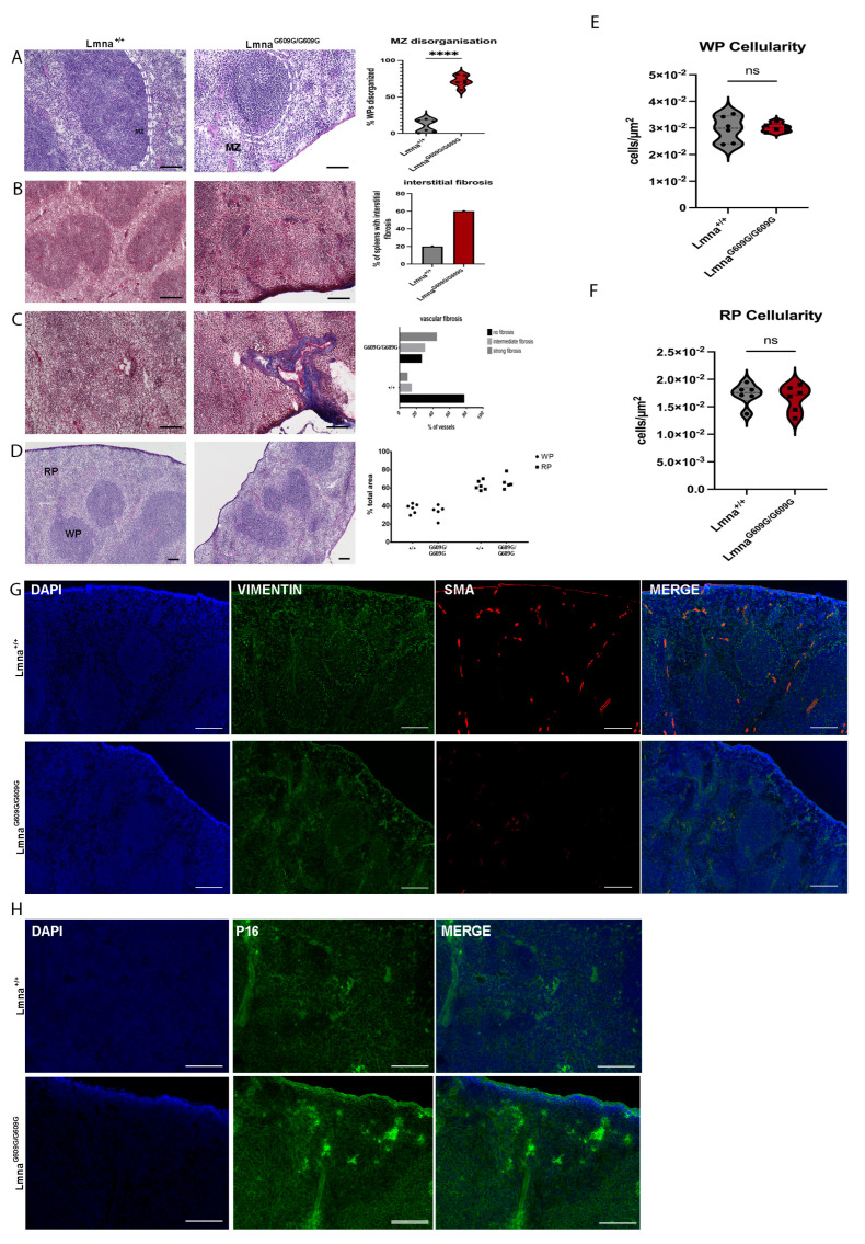
Figure 7.
Histological and Immunofluorescent characterization of splenic pathology in LmnaG609G/G609G mice compared to wildtype C57B/6 mice using a Keyence BZ-X810 microscope and an Axio Imager D2. (WP = white pulp, RP = red pulp, MZ and dashed white lines indicating marginal zone, C = splenic capsule). Histopathological and immunofluorescent staining at 10× and 40× magnification using the Keyence BZ-X810 microscope: (A) H&E staining of spleen showing WP, RP, and spleen capsule. Mutant animals exhibit MZ disorganization at the border between RP and WP (n = 6; **** p < 0.00005; scale bar = 100 µm). (B) Masson’s Trichrome staining indicating elevated interstitial and vascular fibrosis in the spleen of LmnaG609G/G609G mice (n = 6; scale bar = 100 µm). (C) Masson’s Trichrome staining indicating increased vascular fibrosis in the spleen (n = 6; scale bar = 100 µm). (D) Ratio of total WP and RP area was compared between mutant and wildtype cohorts showing no difference. (E) Comparing WP cellularity between LmnaG609G/G609G and wildtype mice. (F) Comparing WP cellularity between LmnaG609G/G609G and wildtype mice. (G) Vimentin (green), αSMA [35], and DAPI staining ECM producing myofibroblasts, vascular smooth muscle cells, and nuclei in the spleen. Vimentin staining reveals a distinct border co-localized with the MZ separating the WP and RP showcasing the structural integrity in Lmna+/+ spleens, while its disorganization is evident in the LmnaG609G/G609G mouse spleen. αSMA was reduced in LmnaG609G/G609G mice. (H) DAPI and p16 staining of spleen tissue. p16 signal is increased in samples of LmnaG609G/G609G mice displaying increased intensity mostly inside the WP region indicating increased senescence.

