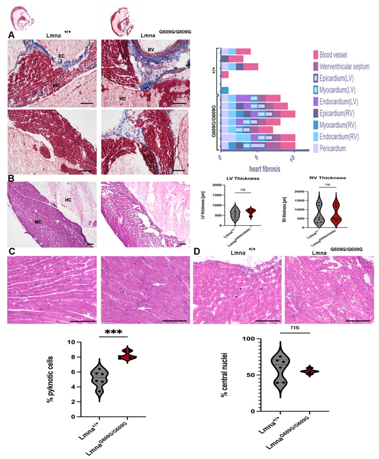
Figure 9.
Histological and Immunofluorescent characterization of cardiac pathology in LmnaG609G/G609G mice compared to wildtype C57B/6 mice (HC = heart chamber; MC = myocardium; EC = endocardium; EPC = epicardium): (A) Masson’s Trichrome staining indicating cardiac fibrosis. Cardiac fibrosis was observed in different cardiac regions (Blood vessel, Interventricular Septum, Epicardium, Endocardium, Myocardium, Pericardium) of Lmna+/+ and LmnaG609G/G609G mice (n = 6 (wt)/n = 7 [25]; scale bar = 100 µm). (B) Left Ventricle cardiac wall thickness was measured (n = 6; p > 0.05 (ns), scale bar = 100 µm). (C) Pyknotic cells are more frequent in LmnaG609G/G609G mice (n = 6; *** p < 0.0005, scale bar = 100 µm). (D) Nuclear position in cardiac myocytes was examined. Peripheral nuclei are marked by a black asterisk, central nuclei are marked by white arrows (n = 6; p > 0.05 (ns), scale bar = 100 µm).

