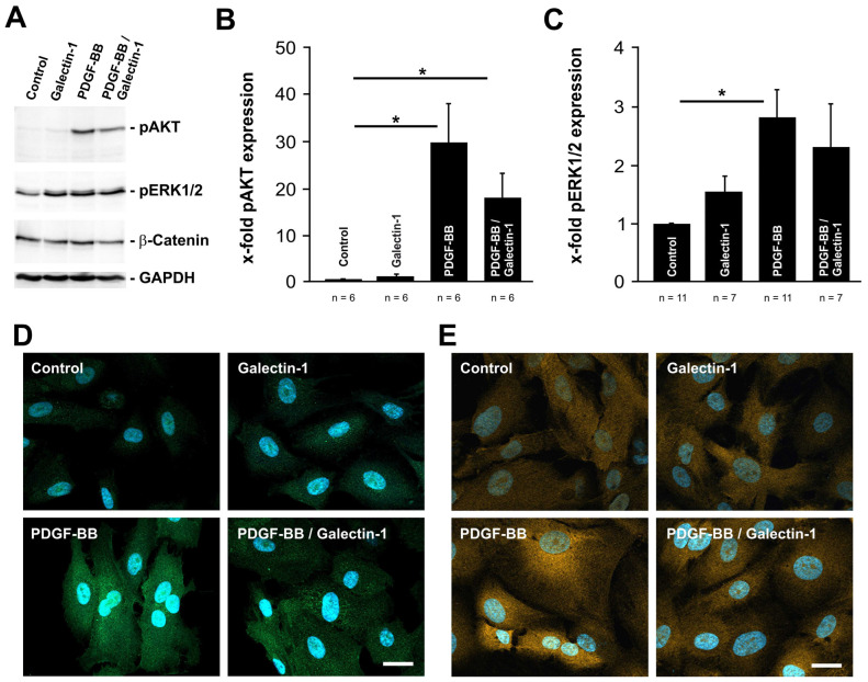
Figure 2.
PDGF-BB-induced AKT and ERK1/2 signaling is reduced by hr-galectin-1. (A–C). Western blot analysis (A) and densitometry (B,C) for pAKT, pERK1/2 and β-catenin of human RPE cells after treatment with 10 µg/mL hr-galectin-1 and/or 20 ng/mL PDGF-BB for 30 min. Mean ± SEM; * p < 0.05. (D,E) Representative immunohistochemical staining against pAKT (D) and pERK1/2 (E) of human RPE cells after incubation with 10 µg/mL hr-galectin-1 and/or 20 ng/mL PDGF-BB for 30 min. Blue, DAPI staining; scale bar, 50 µm.

