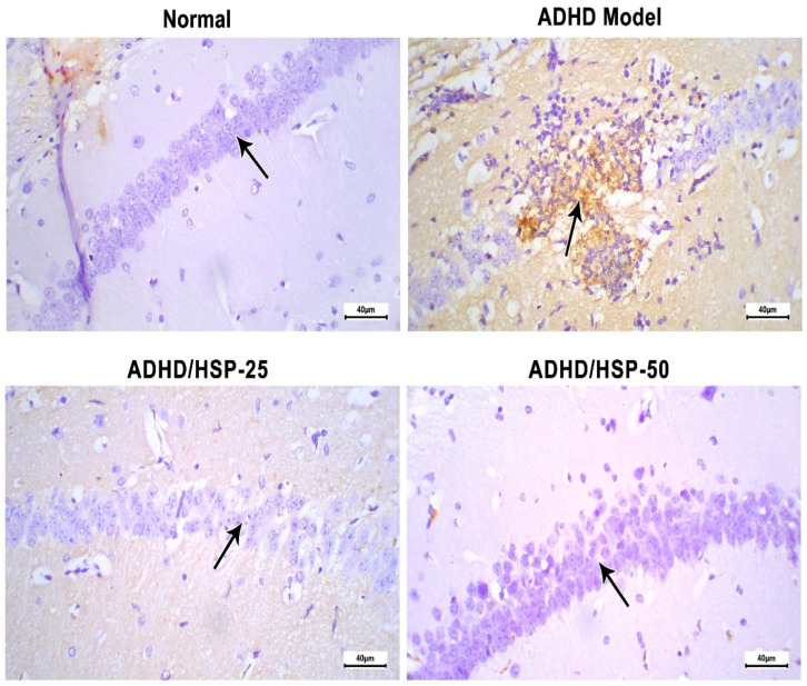
Figure 10.
Histopathological images of hippocampal sections immune-stained for IL-1β. Control group: Mice fed on the normal diet show fairly negative staining and normal arrangement with prominent nuclei. ADHD model group: An image shows degenerated neurons with strong immunostaining in the CA1 hippocampal region. ADHD/HSP-25 and ADHD-50 groups: Arrow indicates the immunostaining in the neurons of the CA1 region (superior region of the Cornu Ammonis). Images show a gradual decrease in immunostaining in the CA1 region. Immunostaining (×400).

