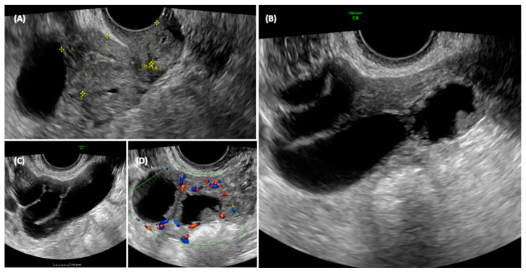
Figure 3.
Ultrasonographic appearance of the pelvis. In the transvaginal scan, the anteverted uterus is visible in the longitudinal section (A), with a size compatible with the patient’s age. At the fundus of the uterus, a cystic formation is visible, closely adhering to the body of the uterus, and posterior to the uterus, the solid component of the formation can be seen. The detail of the cyst formation is visible in images (B,C). A unilocular cyst formation with incomplete septa is observed. In image (B) it appears that there is tissue protruding into the lumen of the cyst, mimicking the characteristic cog-wheel sign typical of inflammatory pathology, but when images are obtained in different planes (C,D) it is clear that this is in fact solid tissue, probably of a neoplastic nature, intensely vascularized.

