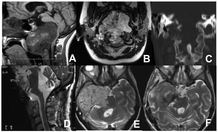
Figure 6.
MRI sagittal (A) and coronal (B) views showing the huge chordoma in the lower third of the clivus, extending to the body of C2, causing posterior displacement of the brainstem, and compressing the nasopharynx. CT scan (C) showing osseous erosion of the occipital condyles, anterior arch of C1, and a portion of the C2 body. Postoperative MR scan after first operation (D). Pre- (E) and postoperative (F) axial MRI scans after the tumor relapse involving the clivus, sphenoid region, right petrous bone, and temporal fossa with severe compression of the brainstem.

