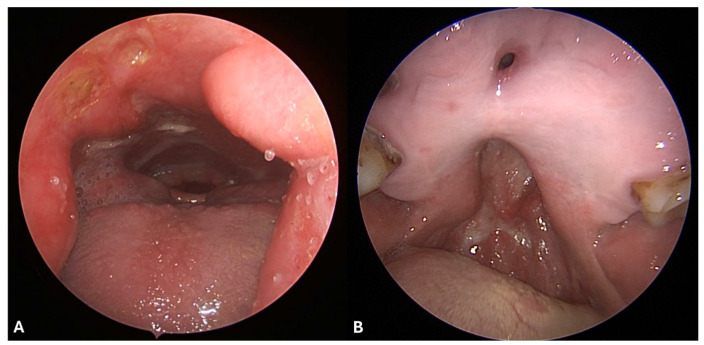
Figure 3.
(A) Nasal endoscopy with a 30-degree endoscope shows a large hard- and soft-palate defect with evidence of oral cavity and laryngopharyngeal structures such as oral tongue, base of the tongue with hypertrophic lingual tonsils, free margin of the epiglottis, laryngeal vestibule, arytenoids, and retrocricoid space. A cluster of three ulcerated lesions of the posterior wall of the nasopharynx is visible. (B) Endoscopic view with a 30-degree endoscope from the oral cavity in the same patient revealed an anterior hard-palate fistula, and a larger hard- and soft-palate defect.

