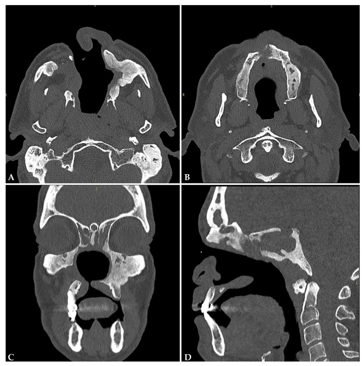
Figure 4.
CT scan of a patient with a history of cocaine abuse, showing in the axial scans (A,B) a complete erosion of the nasal septum and bony limits of the paranasal sinuses, with right paralatero–nasal fistula and hyperostosis of the residual left maxillary-sinus bony walls. This is more evident in the coronal scan (C), where an oronasal fistulation of the hard palate is visible. The ethmoidal cells are obliterated, as well as the frontal and sphenoid sinuses (D).

