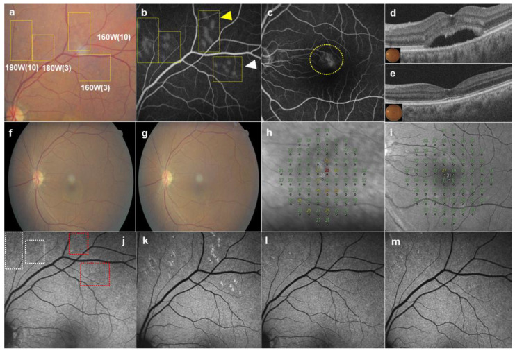
Figure 1.
A 58-year-old female patient presented with a 12-month history of subretinal fluid (SRF) in the left eye. (a) By 1 h after irradiation, 20 test spots (yellow boxes) were barely visible on color fundus photography (CFP). Test spots made with three micropulses at 160 W of pulse energy were less barely visible than those produced with 10 micropulses at 160 W. (b) All barely visible test spots showed leakages on fundus fluorescein angiography (FFA). Test spots produced with three micropulses at 160 W of pulse energy (white arrowhead) showed less leakage than those made with 10 micropulses at 160 W (yellow arrowhead). (c) Thirty-five selective retina therapy (SRT) spots, at one-spot spacing, were produced by 3 micropulses (160 W) in the leakage area (yellow circle) on FFA. (d) SRF was observed on optical coherence tomography (OCT) at baseline. (e) SRF was resolved completely at 6 months post-SRT. No SRT spots were visible on CFP at baseline (f) and at 6 months post-SRT (g). (h) The mean deviation (MD) of microperimetry improved from −1.42 dB at baseline to +0.15 dB (i) at 6 months. (j) Hypoautofluorescent changes at 1 h post-SRT were observed at the test spots produced by 180 W micropulses (white box), but no change was seen at spots produced by 160 W micropulses (red box). (k) Autofluorescence of test spots had increased by 1 month post-SRT, but was decreased at 3 months (l) and 6 months (m) post-SRT. No scotomatous change was observed in the SRT-treated area on microperimetry.

