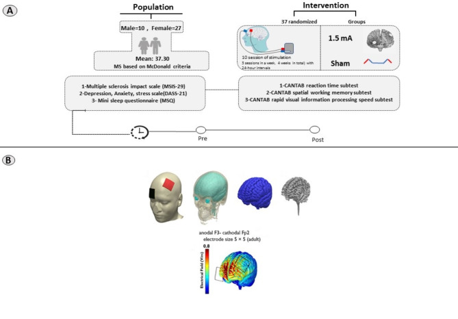
Fig. 2.
(A) This study was a randomized, double-blind trial. Participants were randomly assigned into two groups: active tDCS and sham tDCS. Both groups underwent pre- and post-intervention assessment of mental health-related variables and cognitive performance with the Cambridge Neuropsychological Test Automated Battery (CANTAB), MS battery. The anodal electrode was positioned over the left dorsolateral prefrontal cortex (DLPFC-F3), while the cathode was placed over the right frontopolar cortex (Fp2). (B) 3D models were utilized to examine the flow of electrical current in the brain following the specified protocol. The MR image was segmented into six tissue types: gray matter (GM), white matter (WM), CSF, skull, scalp, and air cavities using SPM8 from the Welcome Trust Center for Neuroimaging, London, UK, with an enhanced tissue probability map. The segmented images were used to create a 3D model with Simpleware software version 5 from Synopsys, Mountain View, CA, incorporating the electrodes and saline-soaked sponges. The distribution of current flow within the brain was then computed using the finite element method in COMSOL Multiphysics software version 5.2 from COMSOL Inc., Burlington, MA. The electric fields were visualized for stimulation intensities of 2.0 mA with an F3 anodal–Fp2 cathodal montage. note: This model illustrates the current flow for 2 mA tDCS for illustrative purposes. The induced electric field of 1.5 mA differs from the 2 mA field

