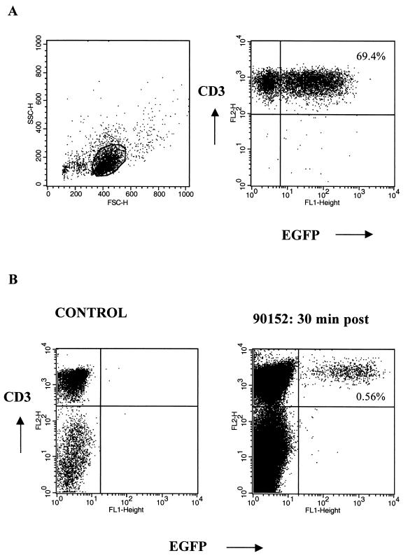
FIG. 2.
(A) Successful gene transfer of M. nemestrina T lymphocytes, demonstrated by representative flow cytometry analysis of EGFP expression in T cells after transduction with the LGSN (PG13) retroviral vector. Gene transfer was accomplished using an optimized transduction protocol including anti-CD3 and anti-CD28 stimulation of T cells. Viable cells were stained with anti-CD3 MAb and analyzed by flow cytometry. Percentages of cells positive for both EGFP and CD3 are as indicated. (B) Transferred EGFP-expressing T cells are detectable by flow cytometry in vivo. PBMC from a control animal (left) and PBMC collected from macaque 90152 at 30 min postinfusion (day 410) (right) were analyzed for expression of both EGFP and CD3. Percentages of cells positive for both EGFP and CD3 are as indicated.

