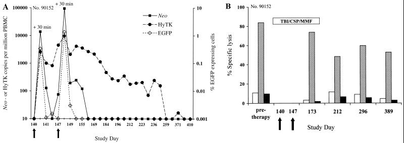
FIG. 6.
(A) Analysis of PBMC for the presence of both EGFP/neo- and HyTK-modified T cells transferred with a transient nonmyeloablative regimen to macaque 90152. The frequency of neo or HyTK sequences was evaluated in samples of DNA derived from PBMC collected before and on the indicated days after infusion, using the real-time PCR assay. The frequency of transferred EGFP-expressing T cells in the same samples of PBMC was assessed by flow cytometry as described in the text. Arrows indicate the days of T-cell infusions. (B) EGFP/neo- and HyTK-specific CTL responses before and after immunosuppressive treatment. Aliquots of PBMC obtained from macaque 90152 at the days indicated on the horizontal axis were stimulated twice with autologous T cells expressing either both the EGFP and neo genes or the HyTK gene and then assayed in a chromium release assay at various E/T ratios for recognition of target cells, either parental (white bars) or transduced to express either both the EGFP and neo genes (hatched bars) or the HyTK gene (black bars). The percent specific lysis of the target cells indicated on the vertical axis is shown for an E/T ratio of 20:1. The presence of EGFP-specific CTL was evaluated in samples of PBMC collected on day 371 instead of day 389. The duration of the treatment is indicated by the horizontal bar, and arrows indicate the days of T-cell infusions.

