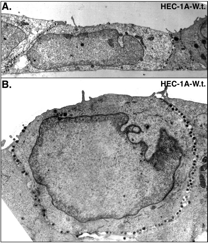
FIG. 1.
Electron micrographs of HEC-1A cells infected with wild-type HSV-1. HEC-1A human epithelial cells were grown to confluence on plastic coverslips and then infected with wild-type HSV-1. After 16 to 18 h, the cells were fixed and processed for electron microscopy. The basal surface is along the bottom of each image.

